
cardiac system pdf
Understanding the cardiac system is crucial, encompassing anatomy, physiology, and devices, as detailed in comprehensive resources like the Handbook.
Essential knowledge covers the heart’s structures, conduction, and vascular networks, vital for grasping cardiovascular function and related disease treatments.
Overview of the Cardiovascular System
The cardiovascular system represents a remarkably complex network, fundamentally responsible for the efficient transport of vital substances throughout the body. This intricate system, as explored in resources like the Handbook of Cardiac Anatomy, Physiology, and Devices, isn’t merely a plumbing network; it’s a dynamic, responsive entity.
At its core lies the heart, a muscular pump propelling blood through a vast array of vessels – arteries, veins, and capillaries. This circulation delivers oxygen, nutrients, hormones, and immune cells to tissues, while simultaneously removing metabolic waste products like carbon dioxide. Biomechanics, as highlighted by Y.C. Fung, plays a critical role in understanding the mechanics of this life-sustaining process.
The system’s efficiency relies on precise coordination, from the heart’s rhythmic contractions to the regulation of blood pressure via structures like the carotid sinuses. Osmosis’s high-yield notes emphasize the importance of grasping these essential anatomical and physiological components. Understanding this overview is foundational for delving into specific cardiac details and potential pathologies.
Importance of Understanding Cardiac Anatomy and Physiology
A robust understanding of cardiac anatomy and physiology is paramount for healthcare professionals and researchers alike. The Handbook of Cardiac Anatomy, Physiology, and Devices underscores this, detailing how knowledge of the heart’s structures directly informs the diagnosis and treatment of cardiovascular diseases.
Grasping the underlying physiological mechanisms – the cardiac cycle, conduction system, and regulation of cardiac output – allows for informed clinical decision-making. Without this foundation, interpreting diagnostic tests, evaluating treatment efficacy, and predicting patient outcomes becomes significantly challenging.
Furthermore, advancements in cardiac research, including the development of animal models and cardiac mapping systems, necessitate a deep comprehension of both anatomy and function. Resources like Osmosis provide visually-rich notes to aid in learning. A solid base enables effective participation in preclinical trials and the application of new technologies, ultimately improving patient care and furthering scientific discovery.
Anatomy of the Heart
The heart’s anatomy encompasses external features, internal chambers, and valves, all crucial for function. Understanding these structures, as detailed in cardiac resources, is key.
External Anatomy of the Heart
The external anatomy of the heart reveals vital clues about its function and relationships with surrounding structures. The heart isn’t a perfectly symmetrical organ; it exhibits a slight leftward rotation, impacting how we view its features. Pericardium, a protective sac, surrounds the heart, containing a small amount of fluid to reduce friction during contractions.
Externally, we observe the coronary sulcus, a groove encircling the heart, marking the separation between the atria and ventricles. Anterior and posterior interventricular sulci delineate the left and right ventricles. These sulci house major coronary arteries and veins, essential for supplying the heart muscle with oxygenated blood and removing waste products.

Auricles, ear-like appendages extending from the atria, increase atrial volume. The apex, the pointed inferior portion of the heart, rests on the diaphragm. Understanding these external landmarks is fundamental to interpreting electrocardiograms and visualizing the heart during imaging procedures. The great vessels – aorta, pulmonary trunk, superior and inferior vena cava, and pulmonary veins – connect to the heart, facilitating blood flow throughout the body.
Internal Anatomy: Chambers and Valves

Internally, the heart comprises four chambers: two atria (right and left) and two ventricles (right and left). The atria are thinner-walled receiving chambers, while the ventricles are muscular pumping chambers. The interatrial septum separates the atria, and the interventricular septum divides the ventricles.
Heart valves ensure unidirectional blood flow. The tricuspid valve lies between the right atrium and right ventricle, while the mitral (bicuspid) valve is positioned between the left atrium and left ventricle. Semilunar valves – pulmonary and aortic – control blood flow exiting the heart. The pulmonary valve guards the exit from the right ventricle into the pulmonary trunk, and the aortic valve regulates flow from the left ventricle into the aorta.
Papillary muscles and chordae tendineae anchor the valve leaflets, preventing backflow during ventricular contraction. These structures work in concert to maintain efficient cardiac circulation. Understanding the precise location and function of each chamber and valve is crucial for diagnosing and treating valvular heart diseases and congenital heart defects;
Cardiac Muscle Tissue (Myocardium)
The myocardium, the heart’s muscular tissue, differs significantly from skeletal muscle. Cardiac muscle cells, or cardiomyocytes, are striated like skeletal muscle but exhibit branching and interconnect via intercalated discs. These discs contain gap junctions, enabling rapid electrical impulse propagation and coordinated contraction.
Unlike skeletal muscle, cardiac muscle is involuntary, meaning it contracts without conscious control. It possesses inherent rhythmicity, generating its own electrical impulses, though this is modulated by the nervous and endocrine systems. The myocardium’s biomechanics are crucial for efficient mass transport, a primary function of the cardiovascular system.
Cardiomyocytes are rich in mitochondria, reflecting their high energy demands. The arrangement of muscle fibers allows for a wringing action during contraction, maximizing ejection fraction. Studying the myocardium, particularly through animal models and cardiac mapping, is vital for understanding heart function and developing treatments for cardiac diseases.

Physiology of the Cardiac System
Cardiac physiology centers on the cycle’s phases, the conduction system’s precision, and output regulation—all essential for mass transport.
Understanding these mechanisms, detailed in resources, is key to grasping cardiovascular function and potential disruptions.
Cardiac Cycle: Phases and Events
The cardiac cycle represents the complete sequence of events occurring during one heartbeat, meticulously orchestrated to ensure efficient blood circulation. This cycle is broadly divided into two primary phases: diastole and systole. Diastole encompasses the relaxation phase, where the ventricles fill with blood, preparing for contraction. This phase includes isovolumetric relaxation, rapid filling, and atrial contraction, contributing to optimal ventricular preload.
Systole, conversely, is the contraction phase, driving blood into the pulmonary artery and aorta. It’s further subdivided into isovolumetric contraction, ventricular ejection, and protodiastole. Crucially, these phases aren’t isolated; they seamlessly transition, governed by pressure gradients and valve function. The coordinated opening and closing of atrioventricular (mitral and tricuspid) and semilunar (aortic and pulmonary) valves are paramount to unidirectional blood flow.
Understanding the timing and interplay of these events is fundamental to assessing cardiac health. Abnormalities in any phase—such as prolonged relaxation or ejection—can indicate underlying cardiovascular dysfunction. Resources like comprehensive handbooks detail these intricacies, providing a foundation for diagnosing and treating cardiac conditions. The cycle’s efficiency directly impacts cardiac output and overall circulatory performance.
Conduction System of the Heart
The heart’s intrinsic conduction system is a specialized network of cells responsible for initiating and coordinating the heartbeat. It begins with the sinoatrial (SA) node, often termed the heart’s natural pacemaker, generating electrical impulses that spread across the atria, causing them to contract. This impulse then reaches the atrioventricular (AV) node, which briefly delays the signal, allowing for complete atrial emptying before ventricular contraction.
From the AV node, the impulse travels down the Bundle of His, dividing into left and right bundle branches, and finally reaching the Purkinje fibers. These fibers rapidly distribute the signal throughout the ventricular myocardium, triggering synchronized contraction. This precise sequence ensures efficient pumping of blood.

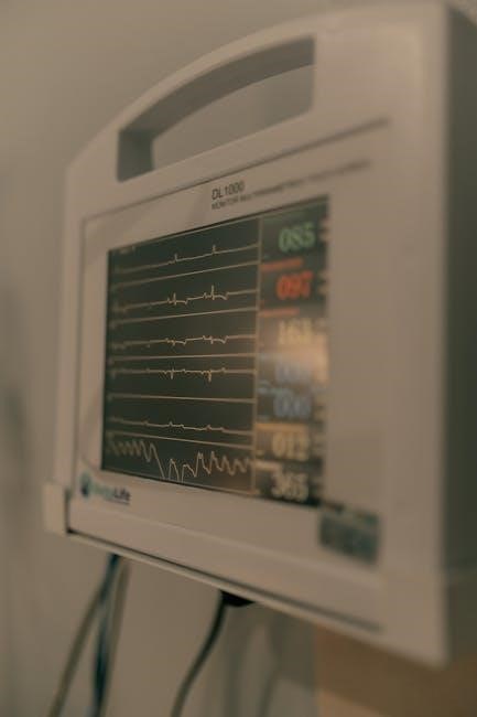
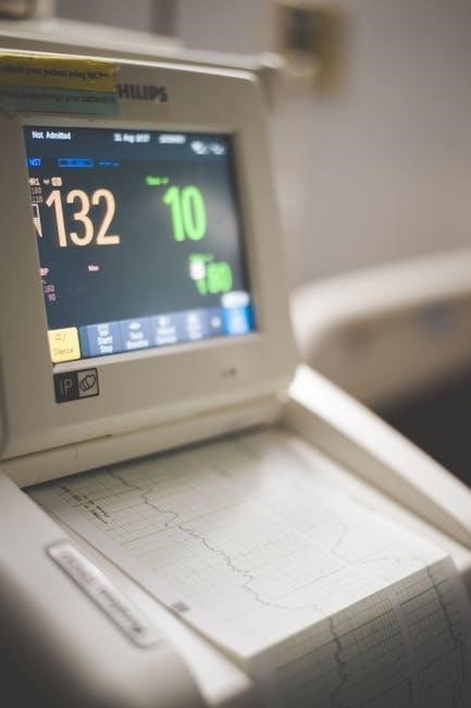
Disruptions within this system—such as blockages or abnormal impulse formation—can lead to arrhythmias. Understanding the conduction pathway is vital for interpreting electrocardiograms (ECGs) and diagnosing cardiac rhythm disturbances. Comprehensive resources, including detailed handbooks, illustrate this system’s complexity and its critical role in maintaining cardiovascular function, emphasizing its importance in overall cardiac health.
Cardiac Output and its Regulation
Cardiac output (CO), the volume of blood pumped by the heart per minute, is a crucial indicator of cardiovascular performance. It’s determined by heart rate (HR) – the number of beats per minute – and stroke volume (SV) – the amount of blood ejected with each beat. Therefore, CO = HR x SV. Maintaining adequate CO is essential for delivering oxygen and nutrients to tissues throughout the body.
Regulation of cardiac output is a complex process involving both intrinsic and extrinsic mechanisms. Intrinsic control relies on the Frank-Starling mechanism, where increased venous return leads to greater ventricular stretch and a stronger contraction. Extrinsic regulation involves the autonomic nervous system and hormones.
The sympathetic nervous system increases HR and contractility, boosting CO, while the parasympathetic nervous system slows HR, reducing CO. Hormones like epinephrine also enhance cardiac function. Furthermore, carotid sinuses, containing baroreceptors, play a role in blood pressure regulation, influencing cardiac output via feedback loops. Understanding these regulatory mechanisms is vital, as detailed in cardiac system resources, for managing cardiovascular health and disease.

Cardiac Vessels
Cardiac vessels, including coronary arteries and veins, are vital for heart function. Carotid sinuses regulate blood pressure, impacting circulation and overall cardiovascular health.
Understanding these networks is key to grasping the cardiac system.
Coronary Arteries and Circulation
Coronary arteries are fundamental to cardiac function, supplying the heart muscle – the myocardium – with oxygenated blood. This vital circulation originates from the aorta, branching into the left and right coronary arteries that encircle the heart. The left coronary artery further divides into the left anterior descending (LAD) and the circumflex artery, while the right coronary artery supplies the right ventricle and portions of the atria.
Efficient circulation within these arteries is paramount; any obstruction can lead to ischemia, potentially causing angina or myocardial infarction. Understanding the anatomical pathways and physiological demands of coronary circulation is crucial for diagnosing and treating cardiovascular diseases. The intricate network ensures continuous nutrient delivery and waste removal, maintaining the heart’s relentless pumping action.
Detailed study of these vessels, often utilizing advanced mapping systems, allows clinicians to pinpoint areas of blockage or narrowing. This knowledge informs interventions like angioplasty or bypass surgery, restoring optimal blood flow and preserving cardiac health. The interplay between coronary anatomy and physiology is a cornerstone of cardiovascular medicine.
Cardiac Veins and Drainage
Cardiac veins play a critical role in returning deoxygenated blood from the myocardium to the right atrium, completing the circulatory loop. These veins branch out from the myocardial capillaries, converging into larger vessels that ultimately drain into the coronary sinus. The coronary sinus, a large vein located on the posterior surface of the heart, empties directly into the right atrium.
Key veins include the great cardiac vein, middle cardiac vein, and small cardiac vein, each following the paths of the coronary arteries. Proper venous drainage is essential for maintaining appropriate cardiac pressure and preventing fluid buildup within the heart muscle. Impairments in venous return can contribute to conditions like heart failure.
Understanding the anatomy of cardiac veins is vital for interpreting diagnostic imaging and planning surgical interventions. Knowledge of venous pathways aids in procedures like cardiac catheterization and the placement of stents. The efficient drainage system ensures the heart functions optimally, removing metabolic waste and maintaining a balanced environment.
Carotid Sinuses and Blood Pressure Regulation
Carotid sinuses, located near the base of the carotid arteries, are crucial chemoreceptor and baroreceptor-rich enlargements that actively participate in blood pressure regulation. These specialized areas detect changes in arterial blood flow and pressure, relaying this information to the central nervous system.
Baroreceptors within the carotid sinuses respond to stretch in the arterial wall, signaling increased blood pressure. This triggers a reflex arc that lowers heart rate and causes vasodilation, reducing blood pressure. Conversely, decreased pressure leads to increased heart rate and vasoconstriction.
This feedback mechanism ensures the body maintains stable blood pressure, vital for adequate tissue perfusion. Dysfunction of the carotid sinuses can lead to conditions like carotid sinus syndrome, characterized by dizziness or fainting. Understanding their role is essential for comprehending cardiovascular control and managing related disorders, as detailed in cardiac system resources.

Advanced Cardiac Topics
Exploring preclinical animal models, cardiac mapping systems, and heart valve disease represents cutting-edge research. These areas advance treatments and deepen our understanding.
Detailed studies enhance clinical trials and improve cardiac care, as found in specialized handbooks and scientific literature.
Animal Models for Cardiac Research
Utilizing animal models is paramount in cardiovascular research, bridging the gap between in vitro studies and human clinical trials. These models allow scientists to investigate complex cardiac physiology and pathophysiology in a controlled environment, mimicking human heart conditions with remarkable accuracy.
Various species, including mice, rats, pigs, and sheep, are employed, each offering unique advantages depending on the research question. For instance, smaller rodents facilitate genetic manipulation, while larger animals more closely resemble human cardiac anatomy and physiology. Researchers can induce conditions like myocardial infarction, heart failure, and arrhythmias to study disease mechanisms and test novel therapeutic interventions.
The Handbook of Cardiac Anatomy, Physiology, and Devices highlights the significance of preclinical animal models, emphasizing their role in evaluating the efficacy and safety of new drugs and devices before human application. Ethical considerations and rigorous experimental design are crucial aspects of this research, ensuring responsible and reliable results that ultimately contribute to improved patient care. Advanced techniques like cardiac mapping are often validated using these models first.
Cardiac Mapping Systems
Cardiac mapping systems represent a significant advancement in electrophysiology, providing detailed visualization of electrical activity within the heart. These technologies are crucial for diagnosing and treating arrhythmias, particularly atrial fibrillation and ventricular tachycardia. They allow physicians to pinpoint the origin and propagation pathways of abnormal heart rhythms with high precision.
Modern systems employ catheters equipped with multiple electrodes to record electrical signals from various points within the heart chambers. This data is then processed and displayed as color-coded maps, revealing areas of abnormal conduction or tissue damage. Non-invasive imaging techniques, such as MRI and CT scans, can be integrated with electrophysiological data to create comprehensive 3D maps of the heart.
As noted in resources like the Handbook of Cardiac Anatomy, Physiology, and Devices, these systems are essential for guiding catheter ablation procedures, where targeted energy delivery eliminates the source of the arrhythmia. Ongoing research focuses on developing even more sophisticated mapping technologies, including high-density arrays and advanced algorithms for improved accuracy and efficiency, ultimately enhancing patient outcomes.
Heart Valve Disease
Heart valve disease encompasses a range of conditions affecting the heart’s four valves – mitral, tricuspid, aortic, and pulmonary. These valves ensure unidirectional blood flow, and dysfunction can lead to stenosis (narrowing) or regurgitation (leakage), disrupting cardiac efficiency. The Handbook of Cardiac Anatomy, Physiology, and Devices highlights the importance of understanding these pathologies.
Stenosis restricts blood flow, forcing the heart to work harder to pump blood through the narrowed opening. Regurgitation allows backflow of blood, reducing forward output and potentially leading to heart failure. Causes include rheumatic fever, congenital defects, age-related degeneration, and infections.
Diagnosis typically involves auscultation (listening for heart murmurs), echocardiography (ultrasound imaging), and potentially cardiac catheterization. Treatment options range from medication to manage symptoms, to valve repair or replacement surgery. Advances in minimally invasive surgical techniques and transcatheter valve therapies are continually improving outcomes for patients with heart valve disease, as detailed in current medical literature.